Discover the role of Indocyanine Green (ICG) fluorescence, medical imaging in early cancer detection and prevention of cancer spread. Learn how this advanced technique enhances tumor visualization, guides surgery, and improves patient outcomes.
Introduction
Cancer remains a leading cause of mortality, often due to late detection and metastasis. Indocyanine Green fluorescence (ICG) imaging is transforming the field of early cancer detection by improving tumor visualization and guiding fluorescence-guided surgery. By identifying sentinel lymph nodes and detecting micro-metastases, ICG helps in preventing the spread of cancer cells, leading to better patient outcomes.”

What is Indocyanine Green (ICG)?
Indocyanine Green (ICG) is a near-infrared (NIR) fluorescent dye that has been widely used in medical imaging. It is injected intravenously and binds to plasma proteins, allowing it to circulate through the body. Due to its high tissue penetration and fluorescence properties, ICG has gained significant attention in oncological applications, particularly for detecting tumors, visualizing lymphatic pathways, and guiding surgical resection.
ICG Fluorescence in Early Cancer Detection
1. Enhanced Tumor Visualization
Traditional imaging techniques such as CT scans and MRIs may not always detect small or early-stage tumors. However, ICG fluorescence imaging highlights cancerous tissues with higher sensitivity and specificity, enabling detection at an early stage. Cancer cells often exhibit increased vascular permeability, allowing ICG to accumulate in tumor tissues and enhance their visibility under near-infrared light.
2. Lymph Node Mapping for Metastasis Detection
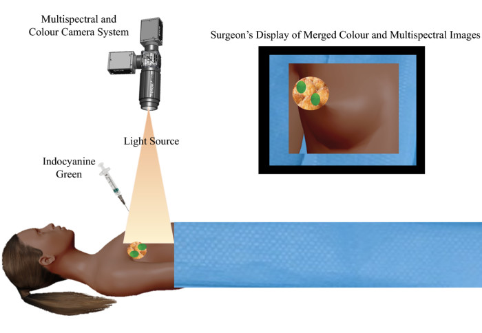
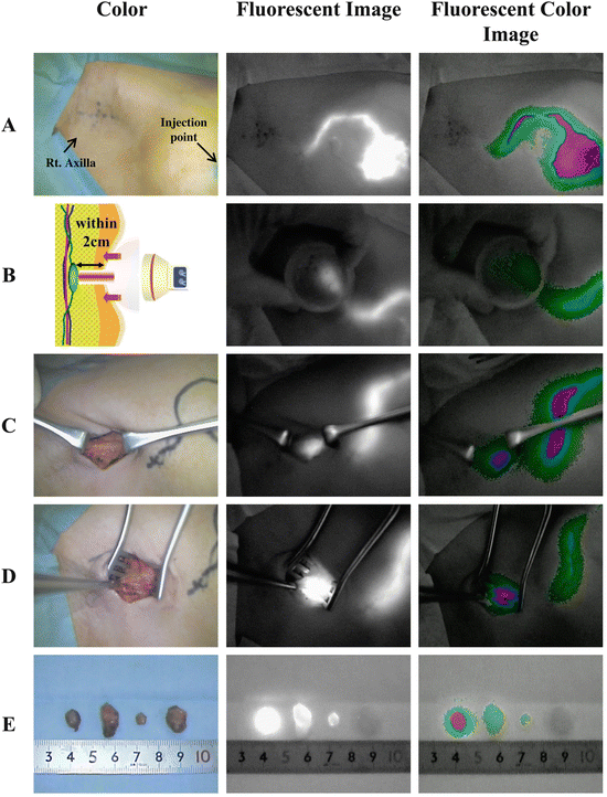
ICG fluorescence plays a crucial role in identifying sentinel lymph nodes (SLNs), which are the first lymph nodes affected if cancer spreads. By injecting ICG near the tumor site, surgeons can track lymphatic drainage pathways and detect potential metastases. This is particularly useful in cancers such as breast cancer, melanoma, and gastric cancer, where early detection of lymph node involvement significantly influences treatment decisions.

3. Margin Assessment During Surgery
One of the biggest challenges in cancer surgery is ensuring complete removal of cancerous tissue while preserving healthy tissue. ICG fluorescence imaging helps surgeons identify tumor margins in real time, reducing the likelihood of residual cancer cells and minimizing the need for additional surgeries.
4. Detection of Micro-Metastases
Traditional imaging techniques may miss micro-metastases, which are tiny clusters of cancer cells that have spread but are too small to be detected by conventional methods. ICG fluorescence can highlight these small lesions, preventing their further spread and improving patient prognosis.
Preventing the Spread of Cancer Cells
ICG fluorescence imaging is not only a diagnostic tool but also plays a critical role in preventing cancer progression:
- Guided Tumor Resection: By helping surgeons distinguish between cancerous and healthy tissues, ICG reduces the risk of incomplete tumor removal, which could otherwise lead to recurrence.
- Real-Time Metastasis Detection: Early identification of metastatic lymph nodes allows for timely intervention, preventing further cancer spread.
- Minimally Invasive Procedures: ICG-guided imaging enables less invasive surgical techniques, reducing patient trauma and speeding up recovery.
Applications of ICG Fluorescence in Various Cancers
- Breast Cancer: Sentinel lymph node biopsy using ICG helps in staging and treatment planning.
- Gastrointestinal Cancers: Enhances detection of liver metastases and tumor margins in colorectal and gastric cancers.
- Lung Cancer: Assists in detecting small pulmonary nodules that may not be visible on traditional imaging.
- Head and Neck Cancers: Improves surgical precision by highlighting tumors in delicate areas.

Challenges and Future Perspectives
While ICG fluorescence imaging has shown immense promise, some challenges remain:
- Limited specificity: ICG accumulates in inflamed or highly vascularized tissues, which can sometimes lead to false positives.
- Standardization issues: Different protocols for ICG administration can affect imaging outcomes.
- Need for advanced imaging systems: Not all hospitals have access to the specialized NIR imaging equipment required for ICG fluorescence detection.
Future research is focusing on ICG-conjugated tumor-targeting agents, which could enhance specificity and further improve early cancer detection.
Conclusion
Indocyanine Green (ICG) fluorescence imaging is revolutionizing cancer diagnostics and treatment. By enabling early tumor detection, precise surgical resection, and real-time metastasis identification, ICG plays a vital role in preventing the spread of cancer cells. As technology advances, integrating ICG fluorescence with AI and molecular imaging techniques may further improve its accuracy and impact, leading to better patient outcomes and higher survival rates.
FAQs on Indocyanine Green (ICG) Fluorescence Imaging
1. What is Indocyanine Green (ICG) fluorescence imaging?
ICG fluorescence imaging is a medical imaging technique that uses Indocyanine Green (ICG), a near-infrared (NIR) fluorescent dye, to visualize blood flow, lymphatic pathways, and tumors. It helps in early cancer detection, surgical guidance, and lymph node mapping.
2. How does ICG fluorescence work in cancer detection?
When injected into the bloodstream, ICG binds to plasma proteins and accumulates in tumors due to their increased vascular permeability. Under near-infrared light, ICG fluoresces, making cancerous tissues and lymph nodes more visible, allowing for earlier and more accurate detection.
3. What types of cancers can be detected using ICG fluorescence?
ICG fluorescence imaging is widely used in detecting and guiding surgery for:
- Breast cancer (sentinel lymph node mapping)
- Gastrointestinal cancers (colorectal, gastric, liver metastases)
- Lung cancer (small pulmonary nodules)
- Head and neck cancers (oral and throat tumors)
- Gynecological cancers (cervical and ovarian cancer)
4. Is ICG fluorescence imaging safe?
Yes, ICG is considered safe and well-tolerated, with a low risk of allergic reactions. However, patients with iodine allergies or liver disease should consult their doctors before undergoing ICG-based imaging.
5. How is ICG administered?
ICG is injected intravenously or near the tumor site, depending on the procedure. For lymph node mapping, it is injected subcutaneously to track lymphatic drainage.
6. What are the advantages of ICG fluorescence in cancer surgery?
- Real-time tumor visualization for precise removal
- Enhanced lymph node detection to assess metastasis
- Better surgical margin identification, reducing recurrence risk
- Minimally invasive, reducing patient trauma and recovery time
7. How does ICG help in preventing the spread of cancer?
By detecting early-stage tumors, metastatic lymph nodes, and micro-metastases, ICG fluorescence imaging allows for timely intervention and targeted treatment, reducing the risk of cancer spreading.
8. Can ICG fluorescence replace traditional imaging methods like MRI or CT scans?
No, ICG fluorescence is complementary to traditional imaging. While it provides real-time, high-resolution imaging, MRI and CT scans are still needed for deep tissue evaluation and comprehensive tumor assessment.
9. Are there any side effects of ICG?
Side effects are rare but may include:
- Mild nausea or dizziness
- Allergic reactions (especially in iodine-sensitive patients)
- Transient liver function changes (in rare cases)
10. Is ICG fluorescence imaging available in all hospitals?
Not all hospitals have near-infrared (NIR) imaging systems required for ICG fluorescence. It is more commonly available in specialized cancer centers and advanced surgical units.
11. How long does the fluorescence effect of ICG last?
ICG fluorescence typically lasts a few minutes to several hours, depending on the dose and location of administration. It is rapidly cleared by the liver and excreted in bile.
12. What is the future of ICG fluorescence imaging in cancer care?
Research is focusing on ICG-conjugated tumor-targeting molecules and AI-assisted imaging to enhance specificity and accuracy. These advancements could further improve early cancer detection and personalized treatment strategies.
